Showing posts with label Kidney Luo-connecting Channel. Show all posts

Friday, November 25, 2022

The Dai Mai and the Lumbar Plexus

What is it about the dai mai that would make a gallbladder channel point (GB 41 zulinqi) have such an influence on it? Or, stated another way, what is it anatomically that connects these? I feel there is a relationship when you look at the neuromyofascial anatomy. 

Dai Mai: Channel Images from A Manual of Acupuncture, by Peter Deadman
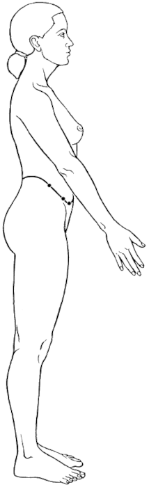
KID Luo

I hear many people say that the transverse abdominis (TrA) is the
 dai mai I think this is not too far off, but it is not technically correct. Interestingly, the TrA is one of the girdling structures of the core and is, in my opinion, a muscle associated with the kidney channel. For ease. I teach that it is part of the kidney sinew channel but actually the kidney luo-connecting channel describes this anatomy the best. The luo channel follows the primary channel in the abdomen. The depth is not described, but it is likely at the depth of the TrA which is the deepest abdominal muscle. The channel then follows as the TrA connects to the diaphragm. which takes it all the way to the central tendon of the diaphragm. The central tendon is a point just below the pericardium, as the pericardium attaches here from above.

The TrA also wraps around and connects with the lateral raphe. This fascial layer then separates into layers of the thoracolumbar fascia and connects with the lumbar multifidi. This is posterior to the spinal column and accessible at the huatuojiaji points. The TrA works with the lumbar multifidi to decompress and stabilize the spine. 


If I have convinced you that the kidney channel relates to the TrA, now we have three things that need to be connected. 1) the dai mai, 2) GB 41, and 3) the kidney channel. Fortunately, there is a really notable link to all of these and this is the lumbar plexus.

The lumbar plexus runs from L1-L4 and has contributions from T12 via the subcostal nerve. The subcostal, iliohypogastric, and ilioinguinal nerves all exit the lumbar plexus, wrap around the abdominal wall, pierce and innervate the lower portions of the abdominals such as the TrA, and obliques. and then become cutaneous.  To me, these nerves are a better representation of the dai mai. 


KID Divergent

Another link of the dai mai and lumbar plexus can be observed. The kidney divergent channel is said to intersect with the dai mai at L2. This channel traverses from the KID 10 region and travels cranially. The pathway, at least, the lower half, follows another nerve of the lumbar plexus which does come from L2. This is the obturator nerve. So, if the kidney divergent channel does have something to do with the obturator nerve (which I think it does) and the dai mai does have something to do with other nerves from the lumbar plexus (which, again, I think it does), then they literally do connect and intersect at L2. 

The final link is that the gallbladder sinew channel is a myofascial plane that runs up the lateral side of the body. It includes the obliques, which are muscles that are innervated from the nerves listed in the lumbar plexus. I think that it is a very plausible that acupuncture to the distal portion of this myofascial plane at GB 41 would communicate mechanical information in the channel, affecting the tone and tension in the obliques, thereby stimulating the nerve coming from the lumbar plexus and innervating these muscles at points such as GB 26 (the motor entry point of the internal obliques) and GB 27 (possibly also a motor entry point of the abdominals). 

I have taught this material at two conferences in 2022. The first was at the Neuroscience Acupuncture Conference and the recording is available through the Neuroscience Acupuncture Conference website.

I also taught a version of this class at the Pacific Symposium, but it was not recorded. 

Finally, the video below looks at activating and strengthening the gallbladder sinew channel, including the obliques. This is to improve the stabilization role of this channel, and balance the left and the right sides and, also the lateral and medial portions of the body. I will be recording another series for this channel soon that has more to do with the rotational role of this channel. While one series will focus more on stabilization and the other on rotation, They each have elements of both stabilization and rotation, You will see some rotation as I get my body into position to activate the channel. When you look at the biomechanics of the pelvic and spinal joints, you see how integrated these to movements are and this helps understand the role of the gallbladder sinew channel for both stabilization and rotation. This starts to highlight the dai mai and its coordinating role for these movements. 

Note: Please support my channel by liking the video, subscribing to the channel, and commenting!


Facebook icon Google Search icon LinkedIn icon Instagram icon YouTube icon

Monday, August 28, 2017

The 'Core' and Channel Relationships

One of the buzzwords in movement and posture-based therapies these days is the 'core.' The exact structures that make up this deep stabilizing region of the body vary depending on the system being studied, but there are some common themes that exist.

The muscles most frequently discussed as 'core muscles' are the deepest of the abdominal muscles. the transverse abdominis; the deep paraspinal muscle group, the multifidi; the pelvic floor muscles; and the diaphragm. These muscles coordinate to stabilize the low spine and pelvis before movement of the legs and arms occurs. This stabilization prevents degeneration of the spinal and pelvic joints. These muscles also work together to align the pelvis and ribcage and properly regulate the position of the spine.

Is there any allusion to these structures in Chinese channel theory? Many channels can be mentioned in this context, notably the extraordinary vessels such as the Daimai, Ren, Chong, and Du. However, it is the Kidney channel that links these structures and can serve as a model to describe this region in TCM terms.

To begin with, the Kidney sinew channel traverses the posteromedial thigh, the pelvic floor and  the spine. I interpret this sinew channel as including the adductor magnus and semimebranosus, which have connections into the pelvic floor muscles (such as the levator ani and obturator internus), which in turn connect to the anterior longitudinal ligament of the spine. The Kidney sinew channel includes some of the stabilizing muscles commonly associated with the core, specifically the pelvic floor.

Another secondary channel of the Kidney serves as a model to integrate function and structure of the core. This is the Kidney Luo connecting channel (Fig. 1). This is how Peter Deadman describes this channel in A Manual of Acupuncture:

  • begins at Dazhong KID-4 on the posterior aspect of the medial malleolus;
  • encircles the heel and enters internally to connect with the Bladder channel;
  • ascends along with the Kidney primary channel from Dazhong KID-4 to a point below the Pericardium where it travels posteriorly to and spreads into the lumbar vertebrae.

     
Fig. 2: Anterior abdominal wall seen from
the posterior view. This shows the
transversalis fascia blending into the
diaphragm. The pericardium would attach
to the central tendon on the superior surface.
Let's break this down and explore the anatomical structures. This secondary channel ascends along the Kidney primary channel. This would include the primary channel abdominal points from KID-11 to KID-22.  This then connects to 'a point below the Pericardium.' All of this can be interpreted to include the transverse abdominis. The fascia of this muscle (especially the transversalis fascia which is posterior to the muscle and anterior to the parietal peritoneum) does link with the diaphragm, and the pericardium of the heart attaches to the central tendon of the diaphragm (Fig. 2). So, there is a continuous plane running up the transverse abdominis to the central tendon just below the Pericardium. 

Fig. 3: Multifidi seen on L
at the Huatuojiaji points.
This channel then travels posteriorly to and spreads into the lumbar vertebrae. Like many of the Yin luo  channels, this channel includes a group of structures which would be on the trajectory of its Yang pair, but at a deeper region of the body. In this case, this would be the lumbar multifidi muscles (Fig. 3). 

I believe the Kidney Luo-connecting channel describes a functional relationship between the transverse abdominis, the lumbar multifidi, the diaphragm and the pelvic floor. These muscles function together to create core support to lift and stabilize the spine. When they are not working well together, there is a collapse that can be seen with a Kidney deficient posture, which overstresses the lumbar spine and contributes to degeneration (See images here, especially Kidney Qi and Jing deficiency). Besides this, when they are not stabilizing the spine and pelvis during activity, there is an increase in wear and tear of these joints. Furthermore, when they are not performing their job adequately, there would be signs and symptoms included in the indications for KID-4, notably, urinary and bowel issues (these muscles are essential to for the pushing type movements necessary for these actions); respiratory issues (tone in these structures regulates intra-abdominal pressure and influences respiration); and lumbar pain and stiffness.

You can work this important region with taiji and qigong exercises. In my own practice, I have been more aware how the timing of these gentle moves facilitates an expansion and contraction of these regions, which both improves tone and elasticity.


But as acupuncturists, we can also wake up this region with direct needling of motor points of these important structures in addition to using distal points such as KID-4. We can also reduce tension in the related Urinary Bladder sinew channel. When deep stabilization of the spine and pelvis is inadequately supported from the muscles associated with the Kidney network, more superficial stabilization occurs, notably from the latissimus dorsi (BL sinew channel) which blends in with the superficial layer of the thoracolumbar fascia (Fig. 4). The thoracolumbar fascia then becomes overly dense and rigid. Reducing tension here can help increase flexibility of the lumbar spine and pelvis and can then allow for adequate training of these deeper Yin muscle counterparts.

Fig. 4: Cross section at lumbar region.


Facebook icon Google Search icon LinkedIn icon Instagram icon YouTube icon