Wednesday, December 21, 2022

Gallbladder Sinew Channel, Side bending and Rotation

Joint mechanics of the spine and pelvis: Coupling Side Bending and Rotation.


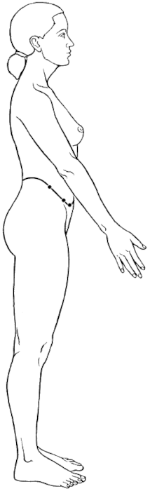
In clinical practice, we see things like an elevated ilium quite frequently. With the elevated ilium, there is almost always some aspect of pelvic torsion. This word can mean different things, but what I mean is that one ilium will be significantly different than the other in terms of anterior and posterior tilts. One side will be significantly more anteriorly tilted or posterior tilted than the other. There will also frequently be rotations in the pelvis and spine.

Why is this? It really has to do with the mechanics of the spine, sacrum and innominate bones. In movements such as walking and running, the body features a combination of side bending and rotation. As the hip flexes and the leading leg reaches out, that innominate bone will roll into a posterior tilt and it will move anterior. Conversely, the opposite innominate will go into an anterior tilt and shift posterior as it follows the back leg in extension. The sacrum will be part of this complex movement and will 'nod' as it rotates and side bends. This nodding is called nutation and counter-nutation and I will not go into detail here other than noting that that the side bending and rotation are coupled in this movement.

The vertebral joints also couple side bending and rotation. For the lumbar and thoracic spine, these movements are coupled in opposite directions. This means that if a vertebra such as T9 right side bends it will also left rotate. This happens at the individual joint level, but you can see the general global spinal pattern in this video where I am demonstrating an exercise called Windmills.


The Gallbladder Sinew Channel performs Side Bending and Rotation.

There are many channels that are involved with these movements. Even the individual muscles of the transversospinalis group (multifidi, rotatores) contribute to the these coupled movement. The attach from inferior transverse processes and reach up to superior spinous processes, pulling the vertebra they insert on into a side bend to that side and a rotation to the opposite side.

The Gallbladder sinew channel supports this and one of its primary actions is to side bend and rotate the torso. Or it stabilizes to prevent excessive side bending and rotation. Either way, it is intimately involved in this movement pattern. Let's look at some key muscles of this channel.

Consider the abdominal obliques. These muscle both side bend the torso, but also rotate the torso. Another example would be the serratus anterior. This muscle abducts the scapula (this is a type of rotation as it rotates around the ribcage) and it also upwardly rotates the scapula (a side bending movement which medially tilts the scapula). We could continue with other examples such as the piriformis, gluteus maximus, gluteus minimus and gluteus medius; and see that all of these muscles have some action that contributes to rotation and side bending. Sometimes these muscles perform side bending and rotation. Other times they stabilize and prevent side bending and rotation. But, their attachments dictate these movements. Here is another video which highlights a training progression to train the stabilization role of this channel. In these exercises, the starting position is side bending and the channel is then engaged to bring the torso back into alignment against gravity. Again, you will see the coupled movement of side bending and rotation.


Facebook icon Google Search icon LinkedIn icon Instagram icon YouTube icon

Sunday, December 11, 2022

Reflections from Cadaver Lab: Week 2 Day 5

Reflecting on my fifth day of week two teaching the 2022 Cadaver Lab.

On the fifth day of lab, we have the donors turned back into a prone position to complete the dissection of the posterior portion of the body. This is actually the first half of the day. For the second half, students are able to finalize any project that they are working with and start exploration to study anything. This week involved exposing the cranial cavity and brain, cutting the vertebral arch to expose the spinal cord and cauda equina, exploring the ligaments of the hip and knee, and dissecting deep into the medial thigh to get to the very deepest layer here. Here are some reflections:

  1. I am teaching, so am back and forth between tables constantly to help advise, help with the dissection, and help identify structures. In some ways this slows down for me during the second half of the day. My colleague that I teach with frequently opens the cranial cavity and highlights structures of the brain. As one would expect, this generates a lot of enthusiasm, and I am usually working to complete a project while he is doing this. This year, I reflected back the muscles of the hip down to the level of the ligaments. This provided a very nice model for these ligaments. I was also able to reflect back to the deepest layer of the groin and expose the obturator externus and also get a very good model of the obturator nerve which could then be followed from the abdominal cavity medial to the psoas and into the medial thigh. This nerve has something to do with the Kidney divergent channel. I feel this is the case because I think that the du mai has something to do with the nerves which exit from the lumbar plexus and wrap around the abdomen to innervate the lower portion of the abdominal muscles such as the internal and external obliques and the transverse abdominis. Specifically, the ilioinguinal, iliohypogastric and subcostal nerve follow the trajectory and describe function of this vessel. I wrote a post about this recently, and you can find this here.

    The Kidney divergent channel is said to intersect with the dai mai at L2, which is exactly what the obturator nerve does. This is why I feel it is part of the Kidney divergent channel, at least the lower half of this secondary channel.

  2. The lumbar plexus is houses between the anterior and posterior layers of the psoas major. We discuss this on an online recorded class available here. I was able to reflect back this anterior layer of the psoas and reveal the lumbar plexus nerves. On this specimen, the psoas was different than I have seen. It had two main grouping of fascicles and almost looked like two separate muscles. This muscle has an interesting fascicle arrangement all of the time, but this was different. I had two distinct branches that then blended with the iliacus muscle. The best I could tell was that the anterior and posterior layer were more distinct from each other than usual, but I was not able to return and figure this out because I got called to other areas for assistance.

  3. Being able to move and palpate a specimen while seeing the structures helps inform what you feel. For instance, mobilizing the hips with just the ligaments holding the hips helps you feel what just the ligaments feel like in this movement. Or doing a varus and valgus stress test with the ligaments exposed helps refine this test. And doing Lachman;s test while watching a torn ACL move gives so much information.


Facebook icon Google Search icon LinkedIn icon Instagram icon YouTube icon

Thursday, December 8, 2022

Reflections from the Cadaver Lab: Week 2 Day 4

Reflecting on my fourth day of week two teaching the 2022 Cadaver Lab.

Each year I teach this cadaver lab, I plan on posting some reflections at the end of each day. I did pretty well this year, posting for days 1-4 of the first week. And then I got off track. This is for two reasons. 1) Dissection lab is very tiring both mentally, but also physically. One is standing the entire day and working over a table, accounting for the physical aspect, but it is also mentally tiring due to the sustained concentration. This is especially true when you are teaching. 2) There is really so much to highlight that at the end of the day it almost makes it too difficult to remember what I was planning to post when I get home. This is made worse by point number one.

Day 4 is the same as last week. It is the day that evisceration occurs and the organs are studied. It is not only this, however. The dissection continues into deeper layers of the anterior neck and extremities so that you can follow myoneurovascular structures from the neck and into both the thoracic cavity but also the upper extremities and you can follow myoneurovascular from the abdominal cavity into the lower extremities. Day 4 is really the culmination of the week up to this point.

Here are some reflections:

  1. There are many things I teach to acupuncturists regarding the channel sinews (jingjin) and their myofascial connections. Reflection of the biceps brachii is a great example of this. With the biceps reflected, you get a great view of the coracobrachialis and the brachialis. The brachialis has two myofascial connections. On the lateral side of the humerus, it has a clear myofascial connection to the deltoids, especially the anterior fibers. Following this path highlights the a deep branch of the Lung sinew channel. However, the brachialis also has a clear myofascial connection to the coracobrachialis which highlights the Pericardium sinew channel. This connection is great in anatomy texts, but much more obvious on a fresh tissue dissection when you can put tension into these myofascial planes. Visually it is apparent, but the tactile portion helps solidify the understanding when considering how injury can affect this plane.

  2. The IT band is really a fascinating structure when you do dissection. It is really almost abstract because, to view this structure, you have to remove the fascia lata (the deep fascia of the thigh) while retaining the IT band. This means you cut an artificial line on the anterior and posterior border and remove the fascia lata off up to this line you created. There is a guideline regarding where you make this line and that is the TFL muscle. The ITB does have some variability in tension from specimen to specimen, but nothing like what you feel when you palpate patient's lateral thighs. There is far more variation with patients. So, all of these tight IT bands really has more to do with the baseline tension in the TFL and/or the underlying vastus lateralis. I think the vastus lateralis is the more likely thing practitioners are palpating. When reflecting the IT band, you follow under the TFL to the ASIS to reflect both together. You have to find the fascial plane between the TFL and the underlying gluteus medius when doing this. It is hard to differentiate. Which is also the case when you palpate and needle these structures on patients. I think many times, clinicians are sensing the gluteus medius and advancing the needle to this muscle when they think they are treating the TFL.

  3. The plantar foot is organized in layers which can be followed in dissection. The superficial layer has the plantar fascia which has a very clear connection to the underlying flexor digitorum brevis, but it also has a clear connection to the adductor hallucis. This is the layer of the Kidney sinew channel. The next layer involves has the flexor digitorum longus, quadratus plantae, and lumbricals. This is the layer of the Liver sinew channel. The final layer includes the tibialis posterior, flexor hallucis and adductor hallucis. This is the layer of the Spleen sinew channel. These layers are well depicted in Netter and other anatomy atlases because the plantar foot is so clearly organized this way in dissection. The channels would follow would also be associated with this order.

  4. I saw a pretty odd anomaly of the psoas major. I will look a bit closer and try to describe tomorrow.


Facebook icon Google Search icon LinkedIn icon Instagram icon YouTube icon

Thursday, December 1, 2022

Reflections from the Cadaver Lab: Week 1 Day 4

Reflecting on my fourth day teaching this year's Cadaver Lab.

Day four of lab is really the point where you start to get into the channels. This happens a bit on day two and three also, but four is much more obvious. We are going more into the neurovascular and myofascial structures, but you are also evicerating. This current class is for Physician Assistant students, so I don't discuss the channels much, but they are always in my mind and here are some anatomical thoughts on the channels that I was observing today.

  1. The greater omentum gives us a view of the Lung channel. Look at the image of this channel in Deadman (or, really any good illustration) and also follow the description and you will see that this channel originates in the region of the stomach and then descends and connects with the large intestine. This is exactly what the greater omentum does. It hangs off of the greater curvature of the stomach and drapes over the abdominal organs. If you lift the bottom of this structure upwards to look at the under surface, you see that this anchors the transverse colon. There is a really interesting physiological thing that connects this structure to the Lungs (capital L meaning that I am discussing the TCM view of the Lung). The greater omentum can move and cover assist when there is infection on the abdominal cavity. It has been described as the abdominal policeman since is has an immune system cells and it surveils the region, mobilizes and covers areas of infection and walls it of with immunologically active tissue. Sounds a bit like wei qi to me!

  2. On the topic of the Lung channel, I was able to get a really great view of the Lung sinew channel. I was demonstrating dissection of the anterior forearm muscles and preparing for reflection of the superficial muscles such as the palmaris longus, flexor capri radialis, and ulnaris. The arm was up overhead to expose the anterior forearm. Removing crosslinks from the ulnar surface of the FCR keeps the lacertus fibrosis (bicipital aponeurosis) intact and you can see such an obvious fascial plane between FCR and the biceps brachii. I pointed out the myofascial plane which interested the students, but I think my acupuncture colleagues would have appreciated it much more.

  3. Another channel I observe when working with a student was the Liver channel and a branch of the Stomach sinew channel. I was helping the student reflect back the quadricep group from the innominate and femur, but keeping them intact as a group and keeping the patellar ligament intact. This requires going medial/under the IT band to remove the vastus lateralis from the linea aspera and other femoral attachments. And it requires going lateral to the medial intermuscular septum of the thigh (the septum between the vastus medialis and adductors) to remove the vastus medialis from the linea aspera and other femoral attachments. Then it requires lifting all of the quadriceps off the femur. When entering into the medial intermuscular septum, you are in the Liver channel. As you follow this space proximal, you end up between the vastus medialis on one side and the distal iliopsoas on the other. You are also in very close proximity to the neurovascular structures in the femoral triangle.

    When you do this on the lateral side, you are cutting the vastus lateralis away from bone. This is a muscle of the Stomach sinew channel. At its most proximal, you need to cut this muscle away from fibrous attachments to the glutes. This is the lateral branch of the stomach sinew channel which is said to connect with the shaoyang channels. It then runs up the gluteus medius and minimus fascia to connect with the lateral raphe, a structure in the thoraculumbar fascia that is the lateral border of the quadratus lumborum and iliocostalis lumborum.


Facebook icon Google Search icon LinkedIn icon Instagram icon YouTube icon

Wednesday, November 30, 2022

Reflections from the Cadaver Lab: Week 1 Day 3

Reflecting on my third day teaching this year's Cadaver Lab.

Things really get moving on day three. The specimens are turned back to supine and we are now differentiating structures so that we can view the musculature and expose neurovascular structures such as those in the femoral triangle, anterior neck, axillary region and down into the extremities. Here are some thoughts from today.

  1. Possibly the biggest roadblock people doing dissection for the first time (really, the first several times) is that, after successfully reflecting the skin and subcutaneous layer, they stop at the hands, feet, pubic area and groin, and the face. This is because these areas are more difficult to dissect and they have a complex anatomy. The problem is that, as you progress deeper, you can't follow the structures fully. For instance, if you don't reflect the skin and adipose at the groin region, you can follow the adductors to their attachments which makes it more difficult to differentiate them. Or, if you don't reflect the skin and subcutaneous tissue on the hands, you can't follow the wrist and finger flexors in the arms to their attachments, This then makes it harder to differentiate the structures which becomes more and more difficult the deep you go.

    This problem becomes apparent on day three. It is always a problem, but I have to say that the class did pretty well with this. I prepared them for the problem with a simple example. I had several shirts on and a hoody. I first took the hoody off, but kept the hood on my head and the sleeve on my right arm. In other words, I didn't completely take it off. Then I proceeded to take of the next shirt. Obviously, since the hoody wasn't removed fully, I couldn't take off the second shirt all the way. Each successive shirt become more problematic.

    I feel a similar problem can occur when working with patients. There can be a tendency to use a needle to release tension in a deep structure. But sometimes we need to unwind tension in the outer, overlying areas to allow the deep structure more room to release.

  2. When differentiating structures, you start to use the scalpel to introduce more movement in the natural planes of movement. This means that you are cutting fascial crosslink between muscles along the natural sliding surfaces. One thing you immediately see is that the muscle becomes loose and floppy which causes it to sag and loose the relationship with other structures. Freeing the crosslinks between the sartorius and underlying quadriceps, for instance, cause it to sag and allows you to move it to see underneath. Seeing this over and over has changed how I work. While we don't want our patients structures to loose integrity and the need these crosslinks between muscles, I do find that I work much more in these fascial spaces, especially with manual work. Encouraging movement in these fascial spaces can have a significant impact on the movement potential of the muscles connected by these crosslinks.


Facebook icon Google Search icon LinkedIn icon Instagram icon YouTube icon

Tuesday, November 29, 2022

Reflections from the Cadaver Lab: Week 1 Day 2

Reflecting on my second day teaching this year's Cadaver Lab.

For description of this series and reflections from Day 1, Click here.

On day two, we flip the donors into a prone position and reflect down to the layer of the deep fascia. For this particular five-day lab, the PA students spend the first half of the day on surface landmarks and suturing. So we only have a half day in the lab doing dissection. Here are some reflections from this day.

  1. I got to practice suturing on the cadaver. This is something that is in the scope for PAs and the dissection lab is a good place from them to start the transition from suture kits to eventually a live person. Obviously, suturing uses a specialized needle. It seems like acupuncturists should be able to do this under our scope. I think as more acupuncturists work in hospitals, this would be a good skill to have and would put them more in demand.

  2. When I show students how to uncover the medial plantar fascia, I describe using the scalpel to cut from the posterior calcaneus and have the blade skim the plantar surface of the calcaneus. The blade then continues directly over the plantar fascia as it removes the skin and adipose. You can use a similar method with palpation of the plantar fascia. You can palpate from the medial side of the foot and feel where the plantar surface of the calcaneus is. As you move more distal past the calcaneus, you would then be just inferior to the depth of the plantar fascia. This gives the depth of where to palpate for plantar fasciitis. However, I often find pain much deeper than this and feel that many times our patients presenting with plantar fasciitis, often are coming in with pain at the quadratus plantae muscle.

  3. The scalpel and the dissection process directly relate to palpation skills. This is not just due to being able to see the structures we work with in dissection, but how you use the blade to uncover layers. One obvious aspect is that, when removing tissue over a muscle, it is best to sweep the blade perpendicular to the muscle fibers. If you sweep the blade parallel to the fibers, it is much more likely that the blade can find the grove between muscle fascicles and go too deep. You can try an experiment yourself that demonstrates this. Sweep your fingers pads of one hand perpendicular across the fingers of your other and notice how the finger pads glide over the fingers. Then do the same in a parallel direction and notice how the pads find the space between fingers and can easily go deep. This is one reason why it is so much easier to feel a muscle when you palpate across the fiber direction. However, when using a blade, you can direct the pressure too deep and start to cut into a muscle or you can come out too superficial and loose the fascial plane. The same can be said with palpation, You have to find the depth and then keep a consistent pressure or you will go too deep or retreat back too superficial and loose the structure you are palpating.

    There is something else that the blade teaches you that expands on this idea of depth. I am going to use an example from day one. When you are reflecting the subcutaneous tissue of the thigh and following from the anterior thigh to the medial thigh, the blade follows the contours of the thigh over the quadriceps. However, you have to anticipate the sartorius muscle or the blade can easily follow this contour and go under the sartorius and cut into it. When you start to see the sartorius, you need to direct the pressure more superficial and over it. This is the same for palpation and I find practitioners struggle with palpating this muscle for the same reason. As you follow the contour of the thigh going in a medial direct, you need to anticipate this muscle and not push your fingers under and deep to it.


Facebook icon Google Search icon LinkedIn icon Instagram icon YouTube icon

Monday, November 28, 2022

Reflections from the Cadaver Lab: Week 1 Day 1

Reflecting on my first day teaching this year's Cadaver Lab.

The first two weeks of every December, I teach two back-to-back five-day dissection labs with the Physician Assistant program at the University of Tampa. When I teach this class each December, I attempt to share some thoughts at the end of each day, but this does not always happen. Dissection takes concentration, and when you are in a teaching role for 24 students, you are tired at the end of the day. This year I will follow through and will keep it simple to make sure I stay on task.

Day one involves some identification of surface anatomy and some time with use of instruments, so it is a shorter day of dissection. We start with reflection of the skin on the anterior portion of the body. At some point, we proceed to a slightly deeper level and reflect the superficial fascia and reach the level of the deep fascia or fascia profunda. Below are some thoughts for the day:

  1. When looking at surface anatomy and noting surgical scars, many students become curious and potentially excited about seeing surgical procedures, especially joint replacements. This is perfectly understandable, especially for healthcare providers. However, I am always struck by how much more complicated and sophisticated the human body is compared to manufactured hip or shoulder. These devices have become much more sophisticated over the years, but they are nothing near the original. Even in a pathological joint, the evidence of the intelligence of the body is on display in the attempt to adapt to injury. I think that the real thing is so complex it can be a bit overwhelming, and when we are new to dissection, we can potentially miss so much that we can't fully appreciate it. You need to see many, many dissections to start to understand the tissue. Having said this, I find it very interesting to see replacements and the effects of other surgical procedures. But, I am more interested in seeing how the body has adapted to this new device or procedure.

  2. At any level of dissection, you are studying movement. I don't need to state the obvious that the donors do not have an intact nervous system, so what can one learn about movement from this process? The answer lies in the fascial sliding surface in the body. Muscles, nerves, blood vessels, lymph vessels, organs, and really all of the body surfaces are connected to one another through fascial crosslinks. When you are introducing your scalpel into these fascial spaces between structures, you are assessing the movement potential between these sliding surfaces and how easily they move in relationship to one another. On day one, when you are removing the skin, you see a difference immediately between different donor bodies. However, there are regions where the skin is much more adhered across the board. The knee, for instance, is like this, especially the medial knee. The adipose is much thinner, and the skin is much more tightly bound to the underlying tissues. While we have ligaments to support the knee and the muscles also play a role, the skin seems to have a supportive role, much like a knee brace.

  3. The adipose varies quite a lot in color, ranging from almost a whitish-pale color to yellow to bright orange. Generally, the paler color is on donors who have much more fluid in the tissue space, whereas the dark orange tends to be on leaner bodies with dryer tissue. For the next two weeks, I am teaching Physician Assistant students, and I have a far easier time discussing these color variations with practitioners of Chinese medicine since we have a system to understand things such as Yin deficiency with deficient heat and Yang deficiency with water overflowing.

  4. The platysma is such a cool muscle!


Facebook icon Google Search icon LinkedIn icon Instagram icon YouTube icon

Friday, November 25, 2022

The Dai Mai and the Lumbar Plexus

What is it about the dai mai that would make a gallbladder channel point (GB 41 zulinqi) have such an influence on it? Or, stated another way, what is it anatomically that connects these? I feel there is a relationship when you look at the neuromyofascial anatomy. 

Dai Mai: Channel Images from A Manual of Acupuncture, by Peter Deadman
KID Luo

I hear many people say that the transverse abdominis (TrA) is the
 dai mai I think this is not too far off, but it is not technically correct. Interestingly, the TrA is one of the girdling structures of the core and is, in my opinion, a muscle associated with the kidney channel. For ease. I teach that it is part of the kidney sinew channel but actually the kidney luo-connecting channel describes this anatomy the best. The luo channel follows the primary channel in the abdomen. The depth is not described, but it is likely at the depth of the TrA which is the deepest abdominal muscle. The channel then follows as the TrA connects to the diaphragm. which takes it all the way to the central tendon of the diaphragm. The central tendon is a point just below the pericardium, as the pericardium attaches here from above.

The TrA also wraps around and connects with the lateral raphe. This fascial layer then separates into layers of the thoracolumbar fascia and connects with the lumbar multifidi. This is posterior to the spinal column and accessible at the huatuojiaji points. The TrA works with the lumbar multifidi to decompress and stabilize the spine. 


If I have convinced you that the kidney channel relates to the TrA, now we have three things that need to be connected. 1) the dai mai, 2) GB 41, and 3) the kidney channel. Fortunately, there is a really notable link to all of these and this is the lumbar plexus.

The lumbar plexus runs from L1-L4 and has contributions from T12 via the subcostal nerve. The subcostal, iliohypogastric, and ilioinguinal nerves all exit the lumbar plexus, wrap around the abdominal wall, pierce and innervate the lower portions of the abdominals such as the TrA, and obliques. and then become cutaneous.  To me, these nerves are a better representation of the dai mai. 


KID Divergent

Another link of the dai mai and lumbar plexus can be observed. The kidney divergent channel is said to intersect with the dai mai at L2. This channel traverses from the KID 10 region and travels cranially. The pathway, at least, the lower half, follows another nerve of the lumbar plexus which does come from L2. This is the obturator nerve. So, if the kidney divergent channel does have something to do with the obturator nerve (which I think it does) and the dai mai does have something to do with other nerves from the lumbar plexus (which, again, I think it does), then they literally do connect and intersect at L2. 

The final link is that the gallbladder sinew channel is a myofascial plane that runs up the lateral side of the body. It includes the obliques, which are muscles that are innervated from the nerves listed in the lumbar plexus. I think that it is a very plausible that acupuncture to the distal portion of this myofascial plane at GB 41 would communicate mechanical information in the channel, affecting the tone and tension in the obliques, thereby stimulating the nerve coming from the lumbar plexus and innervating these muscles at points such as GB 26 (the motor entry point of the internal obliques) and GB 27 (possibly also a motor entry point of the abdominals). 

I have taught this material at two conferences in 2022. The first was at the Neuroscience Acupuncture Conference and the recording is available through the Neuroscience Acupuncture Conference website.

I also taught a version of this class at the Pacific Symposium, but it was not recorded. 

Finally, the video below looks at activating and strengthening the gallbladder sinew channel, including the obliques. This is to improve the stabilization role of this channel, and balance the left and the right sides and, also the lateral and medial portions of the body. I will be recording another series for this channel soon that has more to do with the rotational role of this channel. While one series will focus more on stabilization and the other on rotation, They each have elements of both stabilization and rotation, You will see some rotation as I get my body into position to activate the channel. When you look at the biomechanics of the pelvic and spinal joints, you see how integrated these to movements are and this helps understand the role of the gallbladder sinew channel for both stabilization and rotation. This starts to highlight the dai mai and its coordinating role for these movements. 

Note: Please support my channel by liking the video, subscribing to the channel, and commenting!


Facebook icon Google Search icon LinkedIn icon Instagram icon YouTube icon