Wednesday, June 21, 2023

Liver Sinew Channel Illustrations and Anatomy

Illustrations of the Liver Sinew Channel

It's said that a picture is worth a thousand words. As an educator and anatomist, I find this is especially true. I have often found that very complex anatomical relationships can be understood much more easily by a very good illustration or image. Cadaver dissection is probably the best way to appreciate these relationships, but here are several images that are most certainly better than 'a thousand words and can help until you can get to a dissection. 

Note: we are working on scheduling a 3 day cadaver lab on the East Coast with Sports Medicine Acupuncture and I will be doing a 1 day in Miami in September 2023 with Michael Corradino.  Also note that I will be speaking on the QL and the LIV jingjin at the upcoming FSOMA conference in August 2023

Now on to the anatomy!
Image from Holism Prints
Classically, the Liver sinew channel terminates at the groin. Above is a wonderful illustration from Jennifer Black at Holism Prints which shows the classical view described in Ch 13 of the Lingshu. Below is my anatomical interpretation of this portion of the channel at the groin which involves the anterior adductors (adductor longus, brevis, pectineus, gracilis). All attach to the pelvis on it's inferior surface. This matches the classical view, but does this sinew channel end there?

Image depicting anatomy of the LIV jingjin

Image Toldt's Atlas of Human Anatomy

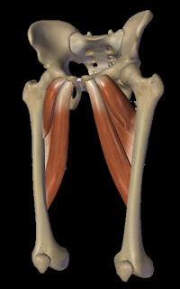
To the left is an image from Toldt's Atlas of Human Anatomy. The patella is facing to the left in this illustration. The perspective is anteromedial which gives a great view of the region of the Liver sinew channel. I produced the labels of the adductor attachments in the intermuscular septum, sometimes referred to as the subsartorial canal (it is deep to the sartorius muscle) and also it is known as Hunter's canal. I teach some manual techniques to open this space and also needle from it angled either anterior into trigger points of the quadriceps or posterior into the adductors. There are blood vessels and nerves running in this space and I feel this canal is where the Liver primary channel runs.

I love this image since is depicts how this myofascial plane continues beyond the groin and into the anterior portion of the pelvis. This entire atlas is amazing!


Toldt's Atlas is from the early 1900s. Let's go back a bit further to 18th century to view an image from Bernhard Siegfried Albinus, a German anatomist, who guided engraver Jan Wandelaar to make a series of fantastic engravings portraying different layers of the body. Here is the deepest layer which beautifully depicts the liver sinew channel all the way up through the anterior pelvis and into the spinal column to the diaphragm!Albinus Anatomy


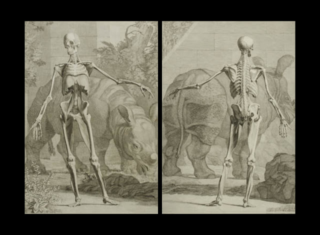
Check out my video that covers this sinew channel from the toes all the way to the diaphragm. Make sure to like the video and let me know if you have any thoughts, questions or considerations in the comment section.


Facebook icon Google Search icon LinkedIn icon Instagram icon YouTube icon

Monday, June 19, 2023

Movement Organized by the Channel Sinews

Channels and Movement Organization

The Channels Organize Movement

If you are an acupuncturist, then you learned a portion of the above image. You learned this as the circadian cycle where energy is at is peak during a 24 hour time period. You also learned various channel correspondences. For instance, the following are internally-externally related channels: LU-LI, ST-SP, HT-SI, KID-UB, P-SJ, LIV-GB. And, finally, you learned the following six divisions: Taiyang (UB-SI), Shaoyang (GB-SJ), Yangming (ST-LI), Taiyin (SP-LU), Shaoyin (KID, HT), Jueyin (LIV-P). You already know that the channels in each of the 3 grouping above share many relationships.  

What you have not been taught, at least in a Chinese Medical Curriculum, is that movement is integrated around these 3 groupings. To fully understand this, you need to dive into the sinew channels (jingjin) and this is exactly what I have been exploring for the last 20+ years. This started with my interested in movement (taiji, qigong, bodyweight calisthenics), developed with my interest in fascial systems such as Anatomy Trains, and continued with exploration on approximately 100 cadaver dissections. This has led to the development of a comprehensive sinew channel model that is taught in the Sports Medicine Acupuncture Certification program where I am a faculty member.

This past Spring 2023, I presented on channels and movement at the first annual Pacific Sports and Orthopedic Acupuncture Symposium (PSOAS). The info below is a summary of this presentation and work I am continuing to develop.

The Channel Sinew Groupings & Functional Movement Patterns

While you can explore what an individual sinew channel does in terms of movement, there is greater benefit in understanding that functional movement patterns are organized by the groupings above. Most movements, especially full body movements  are too complex to analyze with a single channel. Take, for instance, something relatively simple such as a pull up. This movement engages many muscles of the back and involves the UB sinew channel. But the external shoulder rotators are also engaged which would be part of the SI sinew channel. And, if your form is good, there is engagement of core structures such as the transverse abdominis, part of the KID sinew channel. This relatively straightforward movement primarily involves expansive movements that lift and propel the body forward. These movements lift the body up (literally, in the image below), but also lift you into an upright posture, and they also stabilize the spine. They are controlled by the Taiyang and Shaoyin channel sinews.


Expansive movements of the Taiyang-Shaoyin channel sinews
Photo by Ruslan Khmelevsky: https://www.pexels.com/photo/muscular-sportsman-doing-pull-up-exercise-in-gym-4608157/

Other movement patterns guided by the Yangming-Shaoyin channels compress and hollow the body. They protract the scapulae, depress the chest, and flex the torso and hip. Hollow body holds, L-sits, a front kick, and many other movements fall into this category. These movements balance the expansive ones described above and there are many exercises that move back and forth between these two groupings. For instance, rowers would move back and forth between these phases; going into a compression so that they have more range as they engage the back, expand and propel forward as they pull the paddle through the water. You have to compress and load the spring before you expand, especially if you want to do it explosively. The spinal wave from White Crane qigong is another example of this ebb and flow and you can watch my video by clicking here

Compressive movements of the Yangming-Taiyin channels
Photo by Andrea Piacquadio: https://www.pexels.com/photo/photo-of-male-gymnast-practicing-on-gymnastic-rings-3763702/

The final grouping involves the Shaoyang and Jueyin channels which execute two very related movement patterns; sidebending and rotation. This involves trunk and neck sidebending, but also abduction and adduction of the limbs, along with rotational movements. A very simple example is in a video below where I am swinging on monkey bars. This exercise illustrates both rotation, but you will also see sidebending if you watch the video; all engagements of the Shaoyang and Jueyin channels. Note: I mention the Gallbladder sinew channel in this video, but all of the channels in this category are involved. I was simplifying since it is hard to describe these types of concepts in a video without being distracting.

Rotational and Sidebending movements of the Shaoyang-Jueyin channels
Photo by Los Muertos Crew: https://www.pexels.com/photo/woman-doing-sports-at-sports-hall-10039561/

Check out my Youtube channel where I will be exploring many of these concepts. If you do visit, especially if you find the information useful, please subscribe, like the videos, and comment on the videos. All of this helps build my channel and allows me to put the time into producing more content. Besides this, I appreciate the comments since it helps be continue to organize this information and we can all learn together!


Facebook icon Google Search icon LinkedIn icon Instagram icon YouTube icon

Monday, June 5, 2023

The Lateral Raphe: An Important Structure of the Low Back

The Lateral Raphe, Quadratus Lumborum and the 2023 FSOMA conference

I will be presenting about the lateral raphe and the quadratus lumborum at the 2023 Florida State Oriental Medical Association )FSOMA) annual conference on Aug. 27. Details are available here. This post and the video below will give a preview of a portion of this presentation. I hope to see you there!

The Lateral Raphe and Low Back Health

Many manual therapists use high velocity low amplitude (HVLA) adjustments (such as a chiropractic or osteopathic adjustment) to move the body frame and reposition joints. I follow more of the structural integration model where I use the fascia as a lever to move and mobilize the body's skeletal framework. The lateral raphe of the low back is one of these important levers that can influence so much of the low back that it is important to understand the anatomy and use this understanding to better mobilize and move the body.

Netter Anatomy Illustration
Netter Image
showing TLF

The lateral raphe is part of the larger thoracolumbar fascia (TLF). The TLF is the diamond shape aponeurosis (wide, flat tendon) seen in musculature illustration of the back. These illustrations don't do it justice since this isn't simply a single layer structure, but is, instead, a multilayer fascial structure with attachments to so many prominent structures of the low back.

John Hull Grundy Illustration
From Human Structure and Shape
by John Hull Grundy

The fascia from the abdominal muscles, primarily the internal obliques and transverse abdominis continue to wrap around to the back to join with the TLF. Specifically, these fascial layers converge into a seem at the lateral edge of the iliocostalis lumborum and the quadratus lumborum. This seem then separates again into two layers with one layer traveling superficial to the erector spinae to connect with the spinous processes of the lumbar vertebrae, while the second layer travels deep to the erector spinae and between the erectors and the quadratus lumborum (so, superficial to the QL) to connect with the transverse processes of the lumbar vertebrae. This seem is the lateral raphe. It has connections to the abdominals all the way to the rectus abdominis; it has connections to the erector spinae and QL, and it has connections to bony landmarks of the lumbar spine and even the deep lumbar multifidi muscles. 


This fascial seem helps integrate and balance pushes and pulls produced by all of these structures while providing a stable support for the muscles to pull on. You want the lateral raphe to be supple and strong so that it can be a structure that supports the spine while allowing the various muscles that attach to it to communicate mechanically with each other. This mechanic communication is how the myofascial knows where they are in space compared to their functional partners. 

Here is a video featuring palpation and giving a brief demo of techniques to affect the lateral raphe and influence low back health. 


The Lateral Raphe and its Sinew Channel Relationships

The lateral raphe is a meeting point in the fascial system. This plays out also when looking at its channel relationships, particularly looking at the sinew channels/ The following jingjin meet at the lateral raphe:

  • Urinary Bladder jingjin - via pull from the erector spinae
  • Stomach jingjin - following the lateral branch that travels up the vastus lateralis and into the gluteus medius and minimus fascia to the TLF and LR
  • Liver jingjin - via the pull from the quadratus lumborum
  • Kidney jingjin - via the pull from the lumbar multifidi
We will explore this more at the FSOMA conference and look at both local treatment and distal points to influence this important structure/


Facebook icon Google Search icon LinkedIn icon Instagram icon YouTube icon